
Views: 10
Research on an alternative to the traditional mammography is conducted at Chalmers University of Technology. Instead of the current, often painful, X-ray examinations, microwaves can be used for medical imaging of breast tissue to, more accurately, detect breast cancer. The technique is particularly useful as it is capable of also detecting tumours in so called dense breast.
Courtesy Chalmers by Yvonne Jonsson: “Our research indicates that microwave technology can be more effective, gentler and easier than the alternatives available today to diagnose breast cancer”, says Andreas Fhager, Associate Professor of Biomedical electromagnetics at the department of Electrical Engineering at Chalmers.
The method has the potential to eventually replace today’s screening, which women in Sweden aged 40-74 years are offered. Also, it would be advantageous to perform imaging examinations using microwaves to follow-up on patients undergoing breast cancer treatment.
Faster and cheaper system on its way

Within a year, the researchers estimate that they will have a prototype ready for evaluation in the lab setting, which can be used for clinical tests on patients. The new equipment consists of more standardised, and thus cheaper, electronic hardware. In parallel, the software is being adjusted to be able to process the image information faster.
”Thereafter, we will be ready to start planning for clinical studies together with medical staff, to verify that the results obtained by microwave tomography correspond to what we expect”, says Andreas Fhager.
Clinical studies using microwave technology to follow up on breast cancer tumours are already underway in the United States under the supervision of Professor Paul Meaney, the world’s leading researcher in microwave technology for breast cancer imaging. In 2015, he was recruited to Chalmers on part-time and now transfers important knowledge to the Swedish project. The system that the Swedish research group is building is based on Paul Meaney’s research. To further develop the method, collaboration is now under way between researchers in both countries.
Hidden tumours can be detected in dense breasts
Recently, it has been noted that dense beast tissue is one of the major risk factors for developing breast cancer, and the density itself makes it more difficult to detect the cancer.
“Microwave technology would be better suited than traditional mammography to find tumours in women with so-called dense breasts”, says Andreas Fhager. “The images we present show a cross-section of the breast in many layers, and no tumour can be hidden behind other glandular tissue. Very small tumours can also be detected.”
The examination is performed while the patient is lying flat on the stomach on a bunk provided with an opening for the breast, which is lowered into a container with liquid underneath the bunk. In the container a number of narrow upright antennas are positioned, surrounding the breast. The antennas are both transmitters and receivers which in turn send faint microwave signals into the breast. The signals are refracted against the breast tissue and tumours, if any is present, and are then received by the antennas capturing the signal. Depending on whether the tissue is healthy or diseased, the microwaves are reflected in different ways. The pattern that the signals form is then analysed by advanced algorithms for image reconstruction.
“The images we get are rich in contrast, which makes it easier for medical staff to distinguish and assess fat tissue, mammary tissue and tumours,” says Andreas Fhager. “This allows cancer diagnoses to be made more efficiently and accurately.”
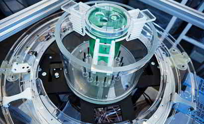
The picture shows the researchers’ prototype of the microwave tomographe. In the transparent container the upright antennas are standing in a circle surrounding the breast. The green coloured container will be filled with tissue mimicking liquid for tests of the imaging system. Some of the connected electronic equipment can be seen below. Photo: Henrik Sandsjö
Enables follow-up examinations
Microwaves, unlike X-rays, emit no ionizing radiation. The research takes into account that microwave tomography is preferable from a radiation point of view, especially to perform repetitive examinations to follow up on how a cancer tumour responds to treatment. Another advantage is that the microwave technology is simple to handle, both for staff and patients. The technology has the prerequisite to become relatively inexpensive. A possible future scenario is small mobile units, that also can be used in developing countries, where healthcare is not fully expanded.
”Many factors indicate that microwave technology has the potential to become a very effective method to fight breast cancer and to reduce mortality caused by the disease”, concludes Andreas Fhager.
What are microwaves?
Microwaves are electromagnetic radiation with shorter wavelength compared to “ordinary” radio waves used for radio communication, but with longer wavelength compared to, for example, visible light and X-rays. This is the same frequency range used, for example, for mobile telephony and wireless networks. The Chalmers researchers use frequencies of about 0.5-3 GHz.
Read more on the research: Biomedical electromagnetics research group
For more information contact:
Andreas Fhager, Associate Professor of Biomedical electromagnetics at the department of Electrical Engineering at Chalmers andreas.fhager@chalmers.se
Related article: Medicine for tomorrow
